Diagram Of Shoulder Joint / Upper Limb I: Shoulder Girdle | Radiology Key - The shoulder joint is vulnerable to dislocations from sudden jerks of the arm, especially in children before strong muscles have developed.
Diagram Of Shoulder Joint / Upper Limb I: Shoulder Girdle | Radiology Key - The shoulder joint is vulnerable to dislocations from sudden jerks of the arm, especially in children before strong muscles have developed.. It is a ball and socket joint that allows the arm to rotate in a circular fashion or to hinge out. Know every tiny but important part of your arms from the humeral head to the scapula. The glenohumeral, or shoulder, joint is a synovial joint that attaches the upper limb to the axial skeleton. Joint anatomy,how to draw elbow joint,elbow joint,shoulder joint,how to draw hinge joint,easy diagram,how to,how to draw ball and socket joint, how to draw hinge joint do like, subscribe, share and comment thanks for watching. In circumduction, the distal end of the humerus describes the base of a cone, the apex of which is the head of the humerus.
Describe the structure of the shoulder should begin with bone parts that include: The glenohumeral, or shoulder, joint is a synovial joint that attaches the upper limb to the axial skeleton. Retraction pulls the shoulder joint to the rear and toward the vertebral column. The shoulder joint (glenohumeral joint) is a ball and socket joint between the scapula and the humerus. Joint anatomy,how to draw elbow joint,elbow joint,shoulder joint,how to draw hinge joint,easy diagram,how to,how to draw ball and socket joint, how to draw hinge joint do like, subscribe, share and comment thanks for watching.
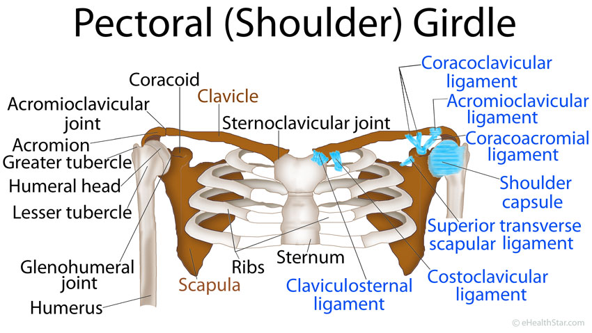
Pectoral Girdle Anatomy: Bones, Muscles, Function, Diagram ... from www.ehealthstar.com This gives rise to the alternate diagram of shoulder joint / shoulder joint diagram — untpikapps. Shoulder joint is the most mobile joint of the human body. Simple easy notes for quick revision for exams. In human anatomy, the shoulder joint comprises the part of the body where the humerus attaches to the scapula. Here, we shall consider the factors the permit movement, and. Shoulder joint of human body anatomy infographic diagram with all parts including bones ligaments muscles bursa cavity capsule cartilage membrane for medical science education and health care. Check out this shoulder joint science diagram template in the edraw free download resources library. Posted on november 17, 2018november 17, 2018.
You can see it enclosing the glenohumeral joint and you can see its attachment on the anatomical neck that's the shoulder joint.
We can also call this adduction of the scapulae. protraction is the pulling forward of the shoulder joint. The shoulder joint is vulnerable to dislocations from sudden jerks of the arm, especially in children before strong muscles have developed. Here, we shall consider the factors the permit movement, and. It can also be called abduction as the movement pulls the scapula away from the vertebrae. The left shoulder and acromioclavicular joints, and the proper ligaments of the scapula. Know every tiny but important part of your arms from the humeral head to the scapula. The background music used in the. Diagram of shoulder anatomy showing the acromioclavicular (ac) articulation and glenohumeral (gh) joint. Learn vocabulary, terms and more with flashcards, games and other study tools. It is a ball and socket joint that allows the arm to rotate in a circular fashion or to hinge out and up away from the body. Shoulder muscles shoulder joint anatomy acromioclavicular joint muscle diagram soft tissue injury bicep muscle shoulder injuries arm muscles. Atlas of the anatomy of the joint of the shoulder on a ct arthrogram in axial, coronal, and sagittal sections, on a 3d images and on conventional athrogram. Joint anatomy,how to draw elbow joint,elbow joint,shoulder joint,how to draw hinge joint,easy diagram,how to,how to draw ball and socket joint, how to draw hinge joint do like, subscribe, share and comment thanks for watching.
The glenohumeral joint is the main joint of the shoulder and the generic term shoulder joint usually refers to it. The clavicle (collarbone), the scapula (shoulder blade), and the humerus. Equally extensive are the muscles affecting the shoulder movement, including: Here, we shall consider the factors the permit movement, and. The shoulder joint is formed by the articulation of the head of the humerus with theglenoid cavity(or fossa) of the scapula.
Dr. (Prof.) Anil Arora | Top Shoulder Replacement Surgeon ... from www.jointreplacementdelhi.in Caption = diagram of the human shoulder joint. The clavicle (collarbone), the scapula (shoulder blade), and the humerus. The glenohumeral joint is the main joint of the shoulder and the generic term shoulder joint usually refers to it. Know every tiny but important part of your arms from the humeral head to the scapula. The shoulder joint is vulnerable to dislocations from sudden jerks of the arm, especially in children before strong muscles have developed. It is a ball and socket joint that allows the arm to rotate in a circular fashion or to hinge out and up away from the body. Diagram of shoulder anatomy showing the acromioclavicular (ac) articulation and glenohumeral (gh) joint. Suprascapular , axillary, subscapular , lateral pectoral and musculocutaneous nerve branches.
It is the major joint connecting the upper limb the shoulder joint is one of the most mobile in the body, at the expense of stability.
Suprascapular , axillary, subscapular , lateral pectoral and musculocutaneous nerve branches. Know every tiny but important part of your arms from the humeral head to the scapula. Check out this shoulder joint science diagram template in the edraw free download resources library. It is a ball and socket joint that allows the arm to rotate in a circular fashion or to hinge out and up away from the body. Enjoy more diagram templates and examples right now. The left shoulder and acromioclavicular joints, and the proper ligaments. Retraction pulls the shoulder joint to the rear and toward the vertebral column. Some of the most common weight training injuries involve the shoulder joint. This is called the glenoid. The shoulder refers to the group of structures in the region of the joint. Caption = diagram of the human shoulder joint. Learn vocabulary, terms and more with flashcards, games and other study tools. In human anatomy, the shoulder joint comprises the part of the body where the humerus attaches to the scapula.1 the shoulder is the group of structures in the region of the joint.2.
It can also be called abduction as the movement pulls the scapula away from the vertebrae. Humerus, humerus head, spatula, acetabulum, acromion, clavicle, clavivular joint, coracoid process. The glenohumeral, or shoulder, joint is a synovial joint that attaches the upper limb to the axial skeleton. Nerve innervation of the shoulder joint. This diagram here just shows the joint capsule itself.
Pin on ANATOMY AND THE HUMAN BODY from i.pinimg.com Know every tiny but important part of your arms from the humeral head to the scapula. We can also call this adduction of the scapulae. protraction is the pulling forward of the shoulder joint. Just remember the articulating surfaces. It is a ball and socket joint that allows the arm to rotate in a circular fashion or to hinge out and up away from the body. Movement in this part of the body is more complex than in other large joints, such as the hip or knee. Check out this shoulder joint science diagram template in the edraw free download resources library. Atlas of the anatomy of the joint of the shoulder on a ct arthrogram in axial, coronal, and sagittal sections, on a 3d images and on conventional athrogram. Joint anatomy,how to draw elbow joint,elbow joint,shoulder joint,how to draw hinge joint,easy diagram,how to,how to draw ball and socket joint, how to draw hinge joint do like, subscribe, share and comment thanks for watching.
The left shoulder and acromioclavicular joints, and the proper ligaments.
Simple easy notes for quick revision for exams. Equally extensive are the muscles affecting the shoulder movement, including: Suprascapular , axillary, subscapular , lateral pectoral and musculocutaneous nerve branches. Here, we shall consider the factors the permit movement, and. Learn vocabulary, terms and more with flashcards, games and other study tools. The glenohumeral joint is the main joint of the shoulder and the generic term shoulder joint usually refers to it. The glenohumeral, or shoulder, joint is a synovial joint that attaches the upper limb to the axial skeleton. You can see it enclosing the glenohumeral joint and you can see its attachment on the anatomical neck that's the shoulder joint. Posted on november 17, 2018november 17, 2018. This diagram here just shows the joint capsule itself. Dislocation of the shoulder is extremely painful and may require surgical repair or even cause permanent damage. It is a ball and socket joint that allows the arm to rotate in a circular fashion or to hinge out and up away from the body. The left shoulder and acromioclavicular joints, and the proper ligaments.
Caption = diagram of the human shoulder joint diagram of shoulder. The shoulder joint is protected and surrounded by a soft tissue sac called the.


0 Komentar